Abstract
In order to achieve its main goal of maximal tumor removal while avoiding postoperative neurologic deficits, neuro-oncological surgery is strongly dependent on image guidance. Among all currently available imaging modalities, MRI provides the best anatomic detail and is highly sensitive for intracranial pathology. However, conventional MRI does not detect the exact location of white matter tracts or areas of cortical activation. This essential information can be obtained non-invasively by means of diffusion tensor MRI (DT-MRI) and functional MRI (fMRI) respectively. Here we present our initial experience with fMRI and DT-MRI for surgical planning and guidance in ten brain tumor cases.
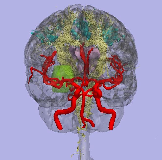
This image displays the cortical surface in transparent gray. Within the brain are shown fMRI activations in teal, arteries in red, tumor in bright green, and white matter fiber tracking in yellow.
| Ion-Florin Talos | talos at bwh.harvard.edu |
| Lauren O'Donnell | lauren at csail.mit.edu |
| Carl-Fredrik Westin | westin at bwh.harvard.edu |