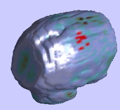
 Functional Magnetic Resonance Imaging (fMRI) assesses brain function by sampling blood flow over time, at thousands of points in the head. Using this imaging modality we can produce "movies" of a subject's brain activity while he is performing a task. The image on the right is a cut-away rendering of one frame of such a movie, in which red indicates high blood flow, and blue, low flow. Our group is investigating ways in which we can use such information to learn more about the interaction among different functional units of the human brain.
One way we're using these data is in learning how brain activity is related to sensory experience and motor function. By doing so, we can use functional brain imaging acquired while a subject is performing a specific task (such as looking a pictures or listening to sound), to localize regions of the brain engaged by that task (such as the visual and auditory areas). This is particularly important information to have during a brain surgery, so that important structures can be avoided.
Finding the relationship between action and brain activity is complicated by the fact that the brain is always engaged in multiple cognitive processes unrelated to the task to which the subject is directing his attention. Furthermore, fMRI is providing only an indirect indication of cognitive function, as blood flow in the brain is also modulated by extrinsic factors such as heart and breathing rate. We use information theoretic techniques distinguish the patterns in a subject's fMRI that relate to what is known about the task he is performing, and thereby ignore confounding influences.
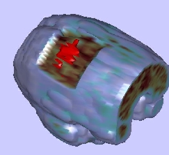
The following movie (avi format, 84Mb) illustrates the process of identifying brain regions related to a subject's activities. It begins with an animation of fMRI values in a subject performing a repetitive motor task, that of repeatedly squeezing a rubber ball. These data are combined with information about how the subject is squeezing the ball, in order to segment related brain regions, which are highlighted in red in the second part of the movie and in the images below. These regions fall primarily in the motor cortex, as is consistent with the nature of the subject's task. Note that this segmentation does not follow directly from inspection of the blood flow, as illustrated in the initial part of the movie where a diveristy of activity is shown throughout the brain.
Junmo Kim, John W. Fisher III, Andy Tsai, Cindy Wible, Alan S. Willsky, William M. Wells III: Incorporating Spatial Priors into an Information Theoretic Approach for fMRI Data Analysis. MICCAI 2000: 62-71 [Postscript]
Andy Tsai,
John W. Fisher III,
Cindy Wible,
William M. Wells III,
Junmo Kim,
Alan S. Willsky:
Analysis of Functional MRI Data Using Mutual Information.
MICCAI 1999: 473-480
Cindy Wible cindy@bwh.harvard.edu
Eric Cosman ercosman@ai.mit.edu
Back to the MIT AI Lab page.