We describe the design of an ophthalmic augmented reality environment. Like the registration system for neurosurgical applications, the goal of this system is to provide the surgeon with an enhanced reality visualization that brings an a priori medical scan into correspondence with the real environment. In this application, real-time photographic retinal imagery through a biomicroscope is registered to previously montaged angiographic retinal image data.
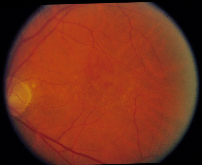
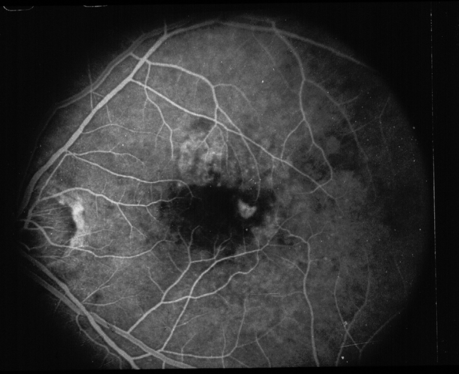
Photographic (left) and fluorescein angiographic (right) image of an eye with age-related macular degeneration. The optic nerve is at the far left of each photograph, with the fovea located centrally. Note that the angiogram conveys additional information regarding areas of leaky blood vessels (for example, the sickle-shaped bright spot in the fovea).
 The sickle shaped structure in the center of the angiographic figure,
above, is a group of leaky blood vessels that need treated by laser
therapy. Without the augmented reality visualization, it is very
difficult to localize this region by just looking at the color video
image, above. Often surgeons have the angiographic data nearby and
attempt mentally register the images to help localize the abnormality.
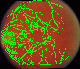
The superposition of the angiographic edges onto the video image
clearly illustrates the position of the treatment area.
The sickle shaped structure in the center of the angiographic figure,
above, is a group of leaky blood vessels that need treated by laser
therapy. Without the augmented reality visualization, it is very
difficult to localize this region by just looking at the color video
image, above. Often surgeons have the angiographic data nearby and
attempt mentally register the images to help localize the abnormality.
The superposition of the angiographic edges onto the video image
clearly illustrates the position of the treatment area.
The registration of these images was computed using the
Hausdorff-distance over translation, rotation, and scale.
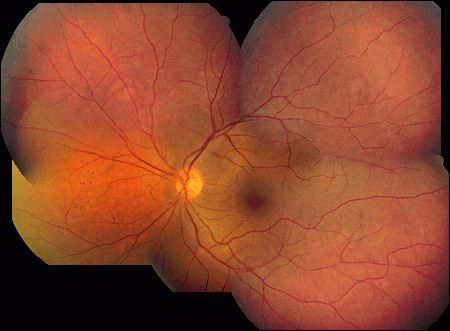
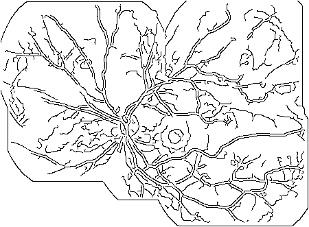 We create a montage of images of the eye taken as the eye looks in
different directions. This registration process is done off-line and
allows for the formation of one complete, coherent dataset to use for
the augmented reality visualization (see figure, below). During a
procedure, the surgeon sees a small region of the retina when looking
through the slit-lamp biomicroscope. The live video image from the
microscope will be registered to the montage dataset. The additional
information in the dataset (for example, the position of leaky blood
vessels) will be superimposed and seen through the slit-lamp
microscope (see figure, above).
We create a montage of images of the eye taken as the eye looks in
different directions. This registration process is done off-line and
allows for the formation of one complete, coherent dataset to use for
the augmented reality visualization (see figure, below). During a
procedure, the surgeon sees a small region of the retina when looking
through the slit-lamp biomicroscope. The live video image from the
microscope will be registered to the montage dataset. The additional
information in the dataset (for example, the position of leaky blood
vessels) will be superimposed and seen through the slit-lamp
microscope (see figure, above).
![]() J.W. Berger,
M.E. Leventon,
N. Hata,
W.M. Wells III,
R. Kikinis.
"Design Considerations for a Computer-Vision-Enabled
Ophthalmic Augmented Reality Environment." In
CVRMED/MRCAS, Grenoble, France, 1997.
[color
postscript 5.0M]
[HTML Version]
J.W. Berger,
M.E. Leventon,
N. Hata,
W.M. Wells III,
R. Kikinis.
"Design Considerations for a Computer-Vision-Enabled
Ophthalmic Augmented Reality Environment." In
CVRMED/MRCAS, Grenoble, France, 1997.
[color
postscript 5.0M]
[HTML Version]
Back to the MIT AI Lab page.