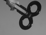
 A related registration and tracking application is the generation of
functional brain mapping using a trans-cranial magnetic stimulation
device. This coil generates magnetic field impulses which stimulate
underlying nerve cells in a focused volume. By measuring responses to
the stimulations and tracking the position of the coil relative to the
MR scan we generate functional maps of the brain in a low-cost,
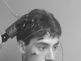
non-invasive, and accurate manner. The LEDs placed on the face in the
image below are used to track the position of the patient during the
procedure, so that the patient's head does not have to be fixed.
Once the coil is positioned as desired, the patient is asked to relax, as the operator counts backwards from five. At the end of the count-down, the magnetic coil is fired and responses from the muscle electrodes are read. A graph of muscle response (in uV) versus time (in ms) is generated for each muscle being mapped. An example graph (waveform) of the forearm muscle appears below.
![]() Each waveform is coded semi-automatically to extract the times
corresponding to the takeoff, peak, trough, and recovery of the
response. The two measures most often used in the analysis of motor
mapping data are latency and amplitude. The latency of the response
is defined as time of the takeoff (in ms) and the amplitude is the
absolute difference between the peak and trough (in uV). When the
magnet is placed directly over the ``hotspot'' of a muscle (or region
of cortex that controls that muscle), the amplitude of the response
should be large and the latency of the response should be small, as
compared to the responses when the coil is place away from the
hotspot.
Each waveform is coded semi-automatically to extract the times
corresponding to the takeoff, peak, trough, and recovery of the
response. The two measures most often used in the analysis of motor
mapping data are latency and amplitude. The latency of the response
is defined as time of the takeoff (in ms) and the amplitude is the
absolute difference between the peak and trough (in uV). When the
magnet is placed directly over the ``hotspot'' of a muscle (or region
of cortex that controls that muscle), the amplitude of the response
should be large and the latency of the response should be small, as
compared to the responses when the coil is place away from the
hotspot.
A typical motor mapping session consists of approximately 120 stimulations at various positions on the head. For each stimulation, for each muscle, we get a latency and amplitude value, and these results are combined to produce motor cortex maps like those that appear below.
The figures below illustrate motor response amplitude maps for four muscles. Green indicates small response. Red corresponds to the largest response. The muscles mapped are (a) index finger, (b) forearm, (c) biceps, and (d) jaw.
The first image below illustrates the responses of the index finger as arrows at each individual stimulation point. Gray arrows correspond no response, and once again, green arrows indicate small responses, while red arrows indicate the the largest response. The second image shows a color coded map of the positions of the minimum latencies and largest responses. Red, yellow, and green are finger, fcr, and biceps respectively, and blue and purple are left oris and right oris respectively.
During a visual suppression mapping session, the volunteer sits in a chair and puts his chin in a chin rest, in front of a monitor. Each visual suppression trial begins with a small dot appearing in the center of the screen. The volunteer is instructed to fixate on the dot, and, when ready, to press the space bar on the computer. At this point, the computer flashes three capital letters in the center of the screen and 90 ms later, fires a train of four pulses on the magnetic coil. If the magnet was directed toward the area of visual cortex, then a momentary blind spot in the visual field will occur. The volunteer is asked to report the three letters that he saw on the monitor. Afterwards, the computer reports the actual three letters. Any discrepancy is considered to be a response and is noted in the results. For visual cortex mapping we treat the responses as binary and thus directly map the points that caused positive suppression in the left and/or right fields of view to the MRI surface points.
In the figures below, red arrows correspond to a suppression in the right visual field, and blue arrows correspond to a suppression in the left visual field. Green arrows indicate no response as the subject correctly reported all three letters.
![]() G. Potts,
L. Gugino,
M.E. Leventon,
W.E.L. Grimson,
R. Kikinis,
W. Cote,
E. Alexander.
J. Anderson,
G.J. Ettinger,
L. Aglio,
M. Shenton,
"Visual Hemifield Mapping Using Transcranial Magnetic Stimulation
Coregistered with Cortical Surfaces Derived from Magnetic
Resonance Images"
J Clin Neurophysiology, 15(4):344-350, 1998.
[html]
G. Potts,
L. Gugino,
M.E. Leventon,
W.E.L. Grimson,
R. Kikinis,
W. Cote,
E. Alexander.
J. Anderson,
G.J. Ettinger,
L. Aglio,
M. Shenton,
"Visual Hemifield Mapping Using Transcranial Magnetic Stimulation
Coregistered with Cortical Surfaces Derived from Magnetic
Resonance Images"
J Clin Neurophysiology, 15(4):344-350, 1998.
[html]
![]() G.J. Ettinger,
M.E. Leventon,
W.E.L. Grimson,
R. Kikinis,
V. Gugino,
W. Cote,
L. Sprung,
L. Aglio,
M. Shenton,
G. Potts,
E. Alexander.
"Experimentation with a Transcranial Magnetic Stimulation
System for Functional Brain Mapping." In
CVRMED/MRCAS, Grenoble, France, 1997.
[color postscript 10.0M]
G.J. Ettinger,
M.E. Leventon,
W.E.L. Grimson,
R. Kikinis,
V. Gugino,
W. Cote,
L. Sprung,
L. Aglio,
M. Shenton,
G. Potts,
E. Alexander.
"Experimentation with a Transcranial Magnetic Stimulation
System for Functional Brain Mapping." In
CVRMED/MRCAS, Grenoble, France, 1997.
[color postscript 10.0M]
![]() G.J. Ettinger,
W.E.L. Grimson,
M.E. Leventon,
R. Kikinis,
V. Gugino,
W. Cote,
M. Karapelou,
L. Aglio,
M. Shenton,
G. Potts,
E. Alexander.
"Non-invasive Functional Brain Mapping Using Registered
Transcranial Magnetic Stimulation." IEEE. Workshop on
Mathematical Methods in Biomedical Image Analysis, San
Francisco, CA, 1996.
[color postscript 6.5M]
[bw postscript 2.7M]
G.J. Ettinger,
W.E.L. Grimson,
M.E. Leventon,
R. Kikinis,
V. Gugino,
W. Cote,
M. Karapelou,
L. Aglio,
M. Shenton,
G. Potts,
E. Alexander.
"Non-invasive Functional Brain Mapping Using Registered
Transcranial Magnetic Stimulation." IEEE. Workshop on
Mathematical Methods in Biomedical Image Analysis, San
Francisco, CA, 1996.
[color postscript 6.5M]
[bw postscript 2.7M]
Back to the MIT AI Lab page.