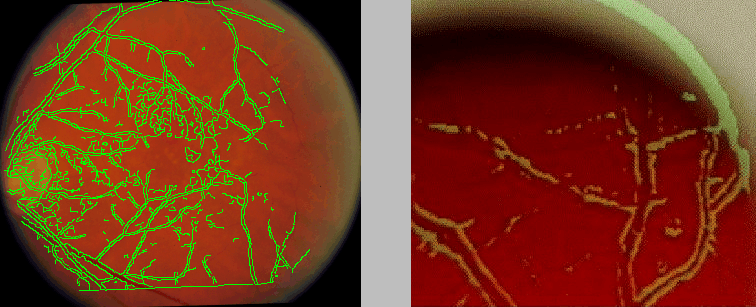
Figure: Image overlay of previously stored edge-detected image of a model eye onto a real-time biomicroscopic image of the model eye. The model eye vessels are not visible in this photograph, but are identified by the image overlay.


Figure: Image overlay of previously stored
edge-detected image of a model eye onto a real-time biomicroscopic
image of the model eye. The model eye vessels are not visible in this
photograph, but are identified by the image overlay.
Figure: Superposition of the edges from
the angiographic image in Figure 2b onto the photographic
image in Figure 1a. The registration was computed using
the Hausdorff-distance over translation, rotation, and scale.
Ultimately, a standard ophthalmic slitlamp biomicroscope will be used together with either a fundus contact lens or a hand-held 60-90 diopter non-contact lens to view the ocular fundus. As an analog to the operating microscope, the slit-lamp biomicroscope is perfectly suited to serve as an imaging and display conduit for AR applications. We are using a modified Zeiss OPM1-DFC operating microscope that permits image overlay of previously stored data on the real-time biomicroscopic image. The microscope is interfaced to a CCD camera, and the image is sent to a frame-grabber and digitizer. The image is processed on a Sun Ultra I workstation where registration calculations are performed. The workstation drives a custom-made high-intensity video display with VGA resolution and adjustable brightness and contrast, and presents images in a false color (e.g. green) to maximize visibility. Algorithm development has utilized both Sun and PC compatible workstations.