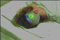
 Diffusion MRI in Neurosurgery
The goal of our group is to develop new algorithms for medical image analysis and visualization of medical imagery, as well as to build vision based systems for surgical navigation and surgical planning. Our group at the MIT CSAIL Lab has been collaborating closely for several years with the Surgical Planning Laboratory of Brigham and Women's Hospital. This page contains links to various projects carried on by the group, as well as the web pages of the group members. The projects are clustered into three categories:
Some colorful movies and powerpoint presentations about certain research projects can also be accessed through this site: PPT .
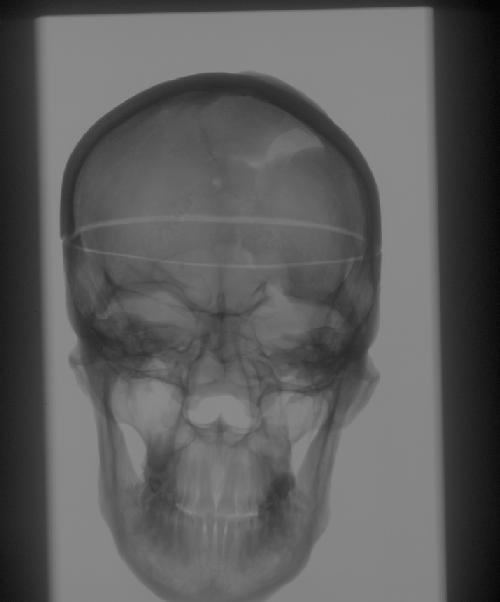
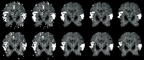
 Epilepsy Surgical Planning
The goal of this project is to accurately detect the epilectic foci in
brain images and plan their resection during brain surgery.
 Anisotropic Modeling of Brain Shift
![]() Intracardiac Surgical Planning
Intracardiac Surgical Planning
 Surgical Navigation
This is a project for enhanced reality visualization of internal anatomical
structures overlaid on live video imagery of patients.
![]() Transcranial Magnetic Stimulation
Transcranial Magnetic Stimulation
![]() Segmentation under Topological constraints: Topology Correction of Digital
Images
Segmentation under Topological constraints: Topology Correction of Digital
Images
![]() A hybrid approach to
the skull stripping problem in MRI
A hybrid approach to
the skull stripping problem in MRI
 Statistical Framework for the Segmentation of Brain Images
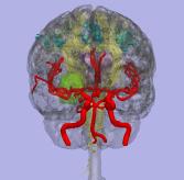
![]() Segmentation of Blood Vessels
in MRA
Segmentation of Blood Vessels
in MRA
![]() Model based segmentation
of Knee MRI
Model based segmentation
of Knee MRI
 A Unified Statistical and Information Theoretic
Framework for Multi-modal Image Registration
A Unified Statistical and Information Theoretic
Framework for Multi-modal Image Registration
![]() Non-rigid registration
of breast MRI
Non-rigid registration
of breast MRI
 Functional Brain Imaging combining DOI and fMRI
 Spatial Regularization of fMRI Activation
Maps
Spatial Regularization of fMRI Activation
Maps
 Exploratory Identification of Cardiac Noise
in fMRI Images
Exploratory Identification of Cardiac Noise
in fMRI Images
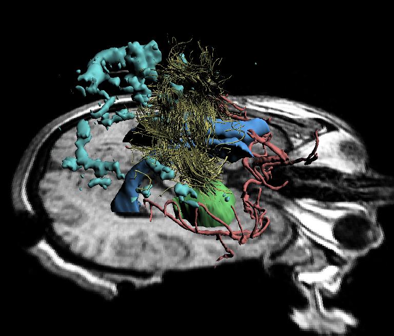
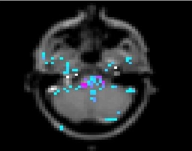
 Neural Connectivity from Diffusion Tensor MRI
![]() Interactive Anatomy Browser
Interactive Anatomy Browser
Anatomy Browser is a tool for visualization and integration of various
kinds of medical information. Its applications include interactive anatomy
atlases, image guided surgery and model driven segmentation.
![]() Patient specific anatomical
models
Patient specific anatomical
models
A collection of patient specific 3D models demonstrating various segmentation
and registration algorithms.
If you are a member of the group and would like to add a project to this page, click here .